Abnormal Chest X-Ray & CT Evaluation Vienna VA
An abnormal chest X-ray or CT scan can raise questions about your lung health, but an abnormal finding does not always mean that a serious condition is present. Chest imaging can reveal many different findings, ranging from temporary changes related to an infection to lung nodules, scarring, inflammation, fluid around the lungs, or signs of chronic lung disease.
At NOVA Pulmonary Critical Care and Sleep Associates, our pulmonary specialists provide abnormal chest X-ray and CT evaluation in Vienna, VA. We review imaging findings in the context of your symptoms, medical history, previous imaging, and other diagnostic testing to help determine whether additional evaluation, monitoring, or treatment may be appropriate.
NOVA Pulmonary Critical Care and Sleep Associates
📍 Vienna Office
124 Park Street SE, Suite 203
Vienna, VA 22180
Map it
📞 571-367-0000
Office Hours:
Mon – Fri: 9:00AM – 5:00PM
Sat & Sun: Closed
What Does an Abnormal Chest X-Ray or CT Scan Mean?
Chest imaging allows physicians to examine the lungs, airways, surrounding tissues, and structures within the chest. An abnormal result simply means that the radiologist or physician identified something that differs from what is typically expected.
Some findings may be minor or related to a previous illness, while others may require additional testing or follow-up imaging.
The significance of an abnormal finding depends on several factors, including its appearance, size, location, whether it has changed over time, your symptoms, and your medical history.
Common Abnormal Findings on Chest Imaging
Chest X-rays and CT scans may identify many different types of abnormalities, including:
✓ Lung nodules or masses
✓ Areas of inflammation or infection
✓ Pneumonia
✓ Pleural effusion or fluid around the lungs
✓ Lung scarring
✓ Pulmonary fibrosis
✓ Emphysema or other signs of COPD
✓ Bronchiectasis
✓ Areas of collapsed lung tissue
✓ Enlarged lymph nodes
✓ Changes involving the airways
✓ Findings that may suggest pulmonary vascular disease
An abnormal imaging result should be interpreted together with your overall clinical picture rather than considered by itself.
When Should You See a Pulmonologist for an Abnormal Chest X-Ray or CT?
Your primary care physician, urgent care provider, emergency department physician, or another specialist may recommend seeing a pulmonologist after an abnormal chest imaging result.
A pulmonary evaluation may be particularly helpful if you have:
✓ A newly discovered lung nodule
✓ Persistent or unexplained abnormalities on imaging
✓ Shortness of breath
✓ A persistent cough
✓ Recurrent respiratory infections
✓ Chest discomfort associated with respiratory symptoms
✓ Abnormal pulmonary function testing
✓ A history of smoking
✓ Known chronic lung disease
✓ Changes compared with previous chest imaging
Even when you do not have respiratory symptoms, certain imaging findings may require monitoring or further evaluation.
Abnormal Chest X-Ray Evaluation in Vienna, VA
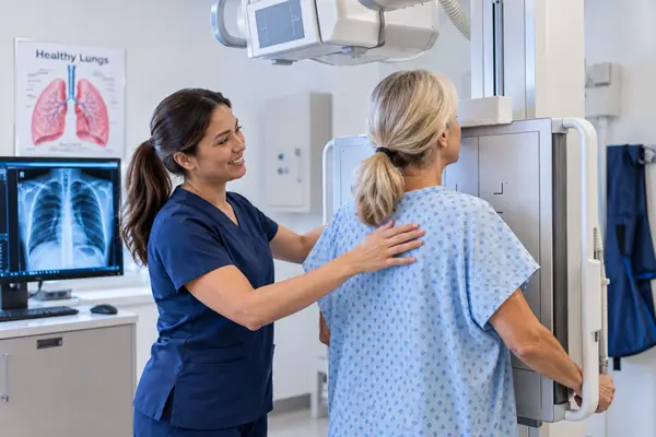
Chest X-rays are commonly used as an initial imaging study when evaluating cough, shortness of breath, chest discomfort, fever, pneumonia, and other respiratory concerns.
Because an X-ray provides a two-dimensional image of the chest, some findings cannot be fully characterized with an X-ray alone. If an abnormality is identified, your physician may recommend comparison with previous X-rays, additional imaging, or a chest CT scan.
The appropriate next step depends on the specific finding and your individual risk factors.
Abnormal Chest CT Evaluation
A chest CT provides more detailed images of the lungs and surrounding structures than a standard chest X-ray. CT imaging can help physicians evaluate lung nodules, scarring, emphysema, bronchiectasis, interstitial lung disease, pleural abnormalities, and other conditions.
If your CT scan has been reported as abnormal, a pulmonologist can review the findings along with your medical history and symptoms to determine what they may mean and whether additional evaluation is necessary.
Evaluation of Lung Nodules
Lung nodules are a relatively common finding on chest CT scans. Many lung nodules are benign and may result from previous infections, inflammation, or scar tissue.
However, some nodules require follow-up because their appearance, size, growth, or other characteristics may warrant closer evaluation.
Your physician may consider factors such as:
✓ Nodule size and appearance
✓ Location within the lung
✓ Whether the nodule is solid or partially solid
✓ Changes compared with previous imaging
✓ Age and medical history
✓ Smoking history
✓ Other individual risk factors
Depending on these factors, monitoring with follow-up imaging or additional diagnostic testing may be recommended.
Scarring and Interstitial Changes on Chest Imaging
Chest imaging may show scarring or changes involving the tissue surrounding the air sacs of the lungs. These findings can sometimes be associated with interstitial lung disease or pulmonary fibrosis.
Not every area of scarring represents progressive lung disease. A pulmonary evaluation can help determine whether the finding appears stable, may be related to a previous infection or injury, or requires additional investigation.
Pulmonary function testing may also be useful for assessing how well the lungs are functioning.
Pleural Effusion on Chest X-Ray or CT
A pleural effusion is an accumulation of fluid in the space between the lung and the chest wall. It may be discovered on a chest X-ray or CT scan performed because of shortness of breath, chest discomfort, cough, or another medical concern.
There are many possible causes of pleural effusion. Evaluation focuses on determining why the fluid developed and whether additional testing or treatment is needed.
Emphysema and COPD Findings on CT
A CT scan may sometimes reveal signs of emphysema even before a patient has been diagnosed with COPD. Patients with these findings may benefit from pulmonary function testing and evaluation of respiratory symptoms.
A pulmonologist can assess whether the imaging findings correspond with changes in lung function and determine whether additional monitoring or treatment may be appropriate.
What to Expect During Your Pulmonary Evaluation
Your pulmonary evaluation generally begins with a review of the radiology report, your symptoms, and your medical history. When available, previous imaging can be especially valuable because it allows physicians to determine whether a finding is new, stable, improving, or changing.
Depending on the abnormality, additional evaluation may include:
✓ Review or comparison of previous chest imaging
✓ Pulmonary function testing
✓ Oxygen level assessment
✓ Follow-up chest X-ray or CT imaging
✓ Laboratory testing when appropriate
✓ Evaluation for infection or inflammatory lung disease
✓ Additional diagnostic procedures when medically indicated
✓ Referral or coordination with other specialists when appropriate
Not every abnormal chest image requires extensive testing. The goal is to determine which findings require additional investigation and which can be appropriately monitored.
Why Comparing Previous Imaging Is Important
One of the most useful parts of evaluating an abnormal chest X-ray or CT scan is comparing it with earlier studies.
A finding that has remained unchanged for an extended period may have a different significance than one that has recently appeared or changed in size or appearance.
If you have previous chest X-rays or CT scans performed at another healthcare facility, making those studies available to your physician may help with your evaluation.
Pulmonary Care in Vienna, Virginia
NOVA Pulmonary Critical Care and Sleep Associates provides comprehensive pulmonary evaluation for patients with abnormal chest imaging and a wide range of respiratory conditions.
Our pulmonary specialists evaluate conditions including lung nodules, pleural effusion, emphysema, COPD, bronchiectasis, pulmonary fibrosis, interstitial lung disease, pulmonary hypertension, recurrent pneumonia, and other pulmonary disorders.
Schedule an Abnormal Chest X-Ray or CT Evaluation in Vienna, VA
If you have received an abnormal chest X-ray or CT result, a pulmonary evaluation can help you better understand the finding and determine whether additional testing or follow-up is needed.
Contact NOVA Pulmonary Critical Care and Sleep Associates to schedule an abnormal chest X-ray or CT evaluation in Vienna, VA.
Directions & Parking Information
Our Vienna office is located on Park Street SE near the Vienna Community Center and historic downtown Vienna.
Free parking available near the building
Elevator access to Suite 203
Easily accessible from Route 123 and I-66
Minutes from Tysons Corner
If you need assistance locating our office, please call and our staff will guide you.
Frequently Asked Questions
Should I be worried about an abnormal chest X-ray?
An abnormal chest X-ray does not necessarily indicate a serious condition. Findings can result from infections, previous illnesses, scarring, fluid, lung nodules, or other causes. Your physician can evaluate the finding in combination with your symptoms and medical history.
Why would I need a CT scan after an abnormal chest X-ray?
A chest CT provides more detailed images than an X-ray and may help physicians better characterize an abnormality that cannot be clearly evaluated on a standard chest X-ray.
What happens if a lung nodule is found on my CT scan?
Many lung nodules are benign. Your physician will consider the nodule’s size, appearance, changes over time, and your individual risk factors to determine whether follow-up imaging or additional evaluation is appropriate.
Can a CT scan detect emphysema?
Yes. Chest CT imaging can show structural changes associated with emphysema. Pulmonary function testing may also be recommended to determine how well the lungs are functioning.
Can a chest X-ray show pleural effusion?
Yes. A chest X-ray can often identify fluid around the lungs. CT imaging or other testing may sometimes be used to further evaluate the amount of fluid and possible underlying cause.
What should I bring to my pulmonary appointment?
Bring your radiology report and, when possible, the actual images from your chest X-ray or CT scan. Previous chest imaging can also be valuable for determining whether a finding has changed over time.
Do all abnormal CT findings require treatment?
No. Some findings require treatment, while others may only need monitoring or no further intervention. The appropriate approach depends on the type of abnormality, your symptoms, medical history, and other risk factors.
Where can I have an abnormal chest X-ray or CT evaluated in Vienna, VA?
NOVA Pulmonary Critical Care and Sleep Associates provides pulmonary evaluation for patients with abnormal chest imaging in Vienna, VA and surrounding Northern Virginia communities.
Office Locations
Conveniently located near you in Dulles, Leesburg, (Loudoun County) and Vienna, (Fairfax County), VA
NOVA Pulmonary – Dulles
24430 Stone Springs Boulevard
Suite 550
Dulles, VA 20166
NOVA Pulmonary – Lansdowne
19415 Deerfield Avenue
Suite 301
Lansdowne, VA 20176
NOVA Pulmonary Critical Care and Sleep Associates is committed to providing excellent care in all aspects of Pulmonary Medicine and Sleep Disorders. With offices located in Lansdowne and Dulles, South Riding, we offer care for the entire Northern Virginia region, serving locations from Chantilly, Fairfax, Vienna, Centreville, Manassas, Gainesville to Reston, Sterling, Leesburg, Ashburn, Brambleton, Purcellville and more.
Meet the team at NOVA Pulmonary Critical Care and Sleep Associates

Dr. Aditya N Dubey, M.D, F.C.C.P. – Founder
Specialty:
Pulmonary, Critical Care and Sleep Medicine
Board Certified by American Board of Internal Medicine in the Subspecialities of Pulmonary Medicine, Critical Care Medicine and Sleep Medicine. Learn more about Dr. Dubey

Dr. Petra Thomas, M.D.
Specialty:
Pulmonary Medicine
Board Certified by American Board of Internal Medicine in the Subspecialities of Pulmonary Medicine. Learn more about Dr. Thomas

Dr. Esha Kapania, M.D.
Specialty:
Pulmonary Medicine
Board Certified by American Board of Internal Medicine in the Subspecialities of Pulmonary Medicine. Learn more about Dr. Kapania

Dr. Arman Murabia, M.D.
Specialty:
Pulmonary, Critical Care and Sleep Medicine
Board Certified by American Board of Internal Medicine in the Subspecialities of Pulmonary Medicine, Critical Care Medicine and Sleep Medicine. Learn more about Dr. Murabia

Dr. Zara Martirosyan, MD
Specialty:
Sleep Medicine & Internal Medicine Specialist
Board-certified physician in both Sleep medicine and Internal medicine. Learn more about Dr. Martirosyan

Rebekah Lee, AGNP-C, Lead APP
Nurse Practitioner. Learn more about Rebekah Lee

Paulos Abebe PA-C
Physician Assistant Learn more about Paulos Abebe PA-C

Katie Cameron, PA
Physician Assistant Learn more about Katie Cameron, PA-C